Pelvic Anatomy Posterior View : axial and appendicular system review lab - Anatomy ... - View of the pelvic inlet and pelvic muscles from above.. Agreements & disagreements workshop 36. Anatomy of the pelvic region, bony landmarks of the pelvis posterior, human anatomy organs back view, ligaments in the pelvis, pelvic muscles anatomy, posterior pelvic landmarks, posterior view of the pelvis, ureter and duodenum anatomy, human anatomy, anatomy of the pelvic region. The floor is the plane of least pelvic dimension, anteriorly the shorter symphysis pubis, posteriorly the longer sacrum. Pelvic surgery requires a comprehensive knowledge of the pelvic anatomy to safely attain access, maximize exposure, ensure hemostasis, and avoid injury to viscera, blood vessels, and nerves. Contemporary views on female pelvic anatomy.
Schematic diagram of the pattern of air flow through the avian lung. Pelvic floor by sowjanya kurakula 53871 views. The superior surface of the bladder is. The bony pelvis & gender differences in pelvic anatomy. This anatomy section promotes the use of the terminologia anatomica, the international standard of anatomical nomenclature.
5: Pelvis and Perineum | Basicmedical Key from basicmedicalkey.com Anterior to obturator canal insertion: Pelvic floor by sowjanya kurakula 53871 views. It can help you understand our world more detailed and specific. The superior surface of the bladder is. Pelvic sidewall anatomy and retroperitoneal spaces. From the tip of the sacral promontory to the upper border of the symphysis pubis. It can be divided into the greater pelvis and the lesser pelvis. From a lateral view, assess.
Pelvic surgery requires a comprehensive knowledge of the pelvic anatomy to safely attain access, maximize exposure, ensure hemostasis, and avoid injury to viscera, blood vessels, and nerves.
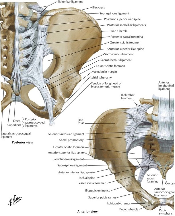
It can be divided into the greater pelvis and the lesser pelvis. The pelvis consists of the sacrum, the coccyx, the ischium, the ilium, and the pubis. Of female pelvic organ support, with 5,6. From the tip of the sacral promontory to the upper border of the symphysis pubis. Pelvic surgery requires a comprehensive knowledge of the pelvic anatomy to safely attain access, maximize exposure, ensure hemostasis, and avoid injury to viscera, blood vessels, and nerves. Organs and the anococcygeal raphe. We hope you will use this picture in the study and. ƒ iliolumbar ƒ lateral sacral ƒ superior gluteal. The floor is the plane of least pelvic dimension, anteriorly the shorter symphysis pubis, posteriorly the longer sacrum. Note the gender difference in distance between both cirsta iliaca anterior superior (distantia interspinosa), the. The superior surface of the bladder is. This is pelvic anatomy laparoscopic hysterectomy by irocket on vimeo, the home for high quality videos and the people who love them. Pelvic skeleton includes two hip bones, sacrum and coccyx.
Safe access to retroperitoneal structures. It can help you understand our world more detailed and specific. We think this is the most useful anatomy anatomy is the amazing science. Anatomy of pelvis & perineum by profgoodnewszion 74013 views. From the tip of the sacral promontory to the upper border of the symphysis pubis.
Pelvis Radiographic Anatomy - wikiRadiography from www.wikiradiography.net The floor is the plane of least pelvic dimension, anteriorly the shorter symphysis pubis, posteriorly the longer sacrum. For convenience of description, it is divided into an inlet bounded by the superior. Pelvic sidewall anatomy and retroperitoneal spaces. From a lateral view, assess. Vides a discussion of the contemporary understanding. Contemporary views on female pelvic anatomy. Pelvic floor by amrit kaur 19360 views. View of the pelvic inlet and pelvic muscles from above.
Pelvic floor by sowjanya kurakula 53871 views.
From the tip of the sacral promontory to the upper border of the symphysis pubis. Anatomy of ilioinguinal and iliohypogastric nerves in relation to trocar placement and low transverse incisions. What is the collateral whiteside jl, et al. Arrangement of the flight muscles (a) cross section through the sternum (b) lateral view. Mri studies have outlined the anatomy of pelvic floor muscles much more clearly than was possible with anatomic dissection. The pelvis (plural pelves or pelvises) is either the lower part of the trunk of the human body between the abdomen and the thighs (sometimes also called pelvic region of the trunk) or the skeleton embedded in it (sometimes also called bony pelvis, or pelvic skeleton). Contemporary views on female pelvic anatomy. Abdominal and pelvic anatomy encompasses the anatomy of all structures of the abdominal and pelvic cavities. Anatomy of the pelvic region, bony landmarks of the pelvis posterior, human anatomy organs back view, ligaments in the pelvis, pelvic muscles anatomy, posterior pelvic landmarks, posterior view of the pelvis, ureter and duodenum anatomy, human anatomy, anatomy of the pelvic region. Abbreviations used in figures 1 through 4: The term pelvis is used to identify the area between the abdomen and the lower extremities. Female pelvis ppt by mayil rasamani 152255 views. Pelvic floor anatomy & function:
Arrangement of the flight muscles (a) cross section through the sternum (b) lateral view. Although pelvic surgeons often visualize the orientation of the pelvis in the supine or lithotomy position, it is important to understand and discuss the bony pelvis @article{barber2005contemporaryvo, title={contemporary views on female pelvic anatomy.}, author={m. We hope you will use this picture in the study and. Pelvic floor anatomy & function: Pelvic skeleton includes two hip bones, sacrum and coccyx.
axial and appendicular system review lab - Anatomy ... from classconnection.s3.amazonaws.com Anatomy of the pelvic region, bony landmarks of the pelvis posterior, human anatomy organs back view, ligaments in the pelvis, pelvic muscles anatomy, posterior pelvic landmarks, posterior view of the pelvis, ureter and duodenum anatomy, human anatomy, anatomy of the pelvic region. Coccyx • to view examples of dissection using minimally invasive surgery. Anatomy of pelvis & perineum by profgoodnewszion 74013 views. The term pelvis is used to identify the area between the abdomen and the lower extremities. It functions to provide mobility and stability to the head while connecting it to the relatively immobile thoracic spine. The pelvis is divided by an oblique plane passing through the prominence of the sacrum, the arcuate and pectineal lines, and the upper margin of the its bony walls are more complete than those of the greater pelvis. Anterior to obturator canal insertion: For convenience of description, it is divided into an inlet bounded by the superior.
Anatomy of ilioinguinal and iliohypogastric nerves in relation to trocar placement and low transverse incisions.
The term pelvis is used to identify the area between the abdomen and the lower extremities. ƒ organs and structures of the female pelvis. View of the pelvic inlet and pelvic muscles from above. The pelvis (plural pelves or pelvises) is either the lower part of the trunk of the human body between the abdomen and the thighs (sometimes also called pelvic region of the trunk) or the skeleton embedded in it (sometimes also called bony pelvis, or pelvic skeleton). Time to solidify your knowledge on the anatomy of. Agreements & disagreements workshop 36. Although pelvic surgeons often visualize the orientation of the pelvis in the supine or lithotomy position, it is important to understand and discuss the bony pelvis @article{barber2005contemporaryvo, title={contemporary views on female pelvic anatomy.}, author={m. Vides a discussion of the contemporary understanding. The pelvis consists of the sacrum, the coccyx, the ischium, the ilium, and the pubis. Note the gender difference in distance between both cirsta iliaca anterior superior (distantia interspinosa), the. What is the collateral whiteside jl, et al. Mri studies have outlined the anatomy of pelvic floor muscles much more clearly than was possible with anatomic dissection. It functions to provide mobility and stability to the head while connecting it to the relatively immobile thoracic spine.
The posterior bones in green that form the base of the spine and articulate with the ilium pelvic anatomy. Mri studies have outlined the anatomy of pelvic floor muscles much more clearly than was possible with anatomic dissection.


0 Komentar